Lachman’s Test
Lachman’s Test is a highly reliable orthopedic examination used to assess the integrity of the anterior cruciate ligament (ACL) in the knee. It is essential for diagnosing ACL tears, which are common in sports and trauma.
How the Test is Performed
-
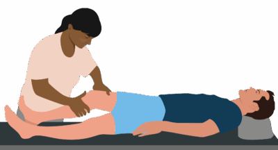
The client lies supine on the table with the knee flexed to about 20-30 degrees.
-
The examiner stabilizes the femur (thigh bone) just above the knee joint with one hand, while the other hand grasps the proximal tibia (shin bone) below the knee.
-
While stabilizing the femur, the examiner applies an anterior force on the tibia to attempt to translate it forward relative to the femur, assessing the amount of movement and the quality of the endpoint.
-
The examiner compares the movement to the unaffected side to detect increased laxity.
-
A positive test is indicated by increased tibial translation with a soft or absent end feel, suggesting ACL rupture.
-
The test can be graded as:
-
Grade 1: mild laxity (2-5 mm difference)
-
Grade 2: moderate laxity (5-10 mm)
-
Grade 3: severe laxity (>10 mm)
-
Clinical Significance
-
A positive Lachman’s Test indicates possible full or partial ACL tear or sprain, which can cause knee instability and dysfunction.
-
It is considered more sensitive and specific than the anterior drawer test, especially in acute injuries.
-
The test is fundamental in early diagnosis and guiding treatment decisions such as conservative care or surgical intervention.
Assessment
-
Use Lachman’s Test when clients report knee instability, giving way, swelling, or trauma mechanisms consistent with ACL injury.
-
Document findings thoroughly, including comparison with the contralateral limb and any accompanying signs (swelling, pain).
Treatment
-
If positive:
-
Avoid aggressive deep tissue work, joint mobilizations, or rapid stretching around the knee in the acute or unstable phase.
-
Focus on gentle soft tissue techniques to surrounding muscles (quadriceps, hamstrings, calf), pain modulation, and support for joint stability.
-
Collaborate with physiotherapists, orthopedic surgeons, and rehabilitation specialists for integrated rather than isolated care.
-
-
Educate clients on proper protection strategies, gradual loading, and bracing as advised medically.
Safety and Referral
-
Refer promptly for further medical evaluation and imaging (MRI) if ACL injury is suspected.
-
Monitor for red flags such as joint effusion, mechanical locking, or neurovascular compromise.