Posterior Drawer Test (Knee)
The Posterior Drawer Test is a clinical orthopedic examination used to assess the integrity of the posterior cruciate ligament (PCL) in the knee. This test is essential for detecting PCL tears or laxity, which can result from trauma (often a direct blow to the front of the shin/tibia).
How is the Test Performed
-
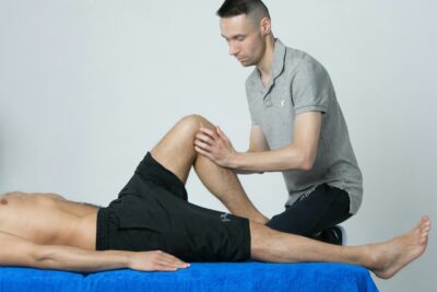
Client position: Supine (lying on their back), hip and knee flexed to 90°, foot flat on the table.
-
The examiner usually sits on the client’s foot (to stabilize) and places hands on either side of the proximal tibia, with thumbs along the tibial plateau at the joint line.
-
The examiner gently pushes the tibia posteriorly (backward) relative to the femur, applying an anterior-to-posterior force.
-
The degree of posterior movement and the quality of the end-feel are compared with the opposite (uninjured) side.
-
A positive test: Excessive posterior translation or absence of a firm end-point compared to the contralateral knee—graded as:
-
Grade 1: 0–5 mm (mild laxity)
-
Grade 2: 6–10 mm (moderate laxity)
-
Grade 3: >10 mm (severe laxity)
-
Clinical Significance
-
A positive Posterior Drawer Test indicates PCL injury, laxity, or tear—PCL is the main restraint to posterior translation of the tibia on the femur.
-
Isolated PCL tears are less common but can result in instability, difficulty with descending stairs/slopes, or persistent knee “giving way”.
-
The test is highly sensitive (~90%) and specific (~99%) for PCL injury, but should be used alongside other clinical findings (e.g., “posterior sag sign,” collateral ligament tests) for accuracy.
Assessment
-
Perform for clients with knee trauma, suspected ligamentous instability, pain, or weakness with stress on the posterior knee.
-
Record the amount of posterior movement, side-to-side difference, end-feel, and associated symptoms for treatment planning and communication.
Treatment
-
If positive:
-
Avoid deep tissue massage, vigorous mobilization, or aggressive stretching around the knee, especially in acute or unstable phases.
-
Focus on gentle soft tissue work for surrounding musculature (hamstrings, gastrocnemius, quadriceps), pain modification, swelling control, and functional stability.
-
Educate clients regarding activity modification and avoid activities that load the PCL (e.g., heavy squatting, downhill running) until cleared by a healthcare provider.
-
Collaborate with physiotherapists, orthopedic surgeons, and rehabilitation teams as indicated.
-
Safety and Referral
-
Refer for further assessment and imaging if significant or acute PCL injury is suspected, instability is marked, or if there are red flag findings.
-
Reassess regularly and modify manual therapy as recovery or post-surgical protocols allow.