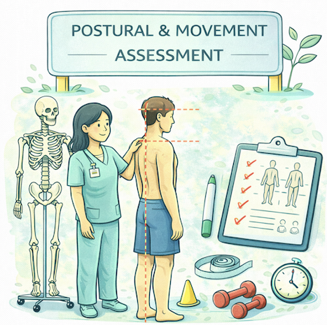
Postural and movement assessment helps massage therapists see how a client’s body is using its tissues, not just how those tissues feel on the table. For students, building this skill turns “where it hurts” into “why it hurts,” so treatment becomes more targeted, safer, and more effective.
Why Posture and Movement Matter
Static posture and dynamic movement give you clues about which muscles are overworking, which are underactive, and where joints may be overloaded. Instead of chasing pain, you start to recognize patterns—like rounded shoulders, anterior pelvic tilt, or limited hip extension—that may contribute to symptoms. This makes your hands‑on work more strategic and improves your ability to give meaningful home care.
 Basic Principles of Observation
You don’t need complex tools to start assessing posture and movement; you need a systematic way of looking.
Key principles:
- Look from multiple views: anterior, posterior, and both lateral sides.
- Compare left and right: height, symmetry, and alignment.
- Observe from global to local: whole body first, then regions (head/neck, shoulders, spine, pelvis, knees, feet).
- Note what is obvious first; refine details later.
Encourage the client to stand and move as naturally as possible. Explain what you’re doing so they don’t “overcorrect” posture to please you.
Simple Postural Observation Table
| View | What to scan for |
|---|---|
| Anterior | Head tilt, shoulder height, rib cage shift, knee valgus/varus, foot arches/toe out |
| Posterior | Scapular winging/elevation, spinal curves, pelvic tilt/shift, Achilles tendon alignment |
| Lateral | Forward head, thoracic kyphosis, lumbar lordosis, pelvic tilt, knee hyperextension |
Common Postural Patterns
Certain patterns show up frequently in clinical practice. Recognizing them helps you link what you see to what you feel during palpation.
Examples:
- Upper crossed pattern: Forward head, rounded shoulders, tight upper trapezius/levator/pectorals, lengthened deep neck flexors and mid‑lower trapezius.
- Lower crossed pattern: Increased lumbar lordosis, anterior pelvic tilt, tight hip flexors and lumbar extensors, lengthened abdominals and gluteals.
- Flat back or sway back: Reduced or shifted lumbar curve, often with hamstring tightness and hip joint strain.
These are patterns, not diagnoses. Use them as hypotheses you will confirm or refine with palpation and client history.
Basic Movement Screens for Massage Therapists
Movement assessment doesn’t have to be complex. A few simple screens can give you useful information before treatment.
Useful tests (within your scope and comfort):
- Cervical and shoulder AROM: Flexion, extension, rotation, abduction—note range, symmetry, and pain.
- Trunk motions: Forward flexion, extension, side flexion—watch how motion spreads through the spine and hips.
- Functional movements:
- Bodyweight squat (depth, knee alignment, trunk control).
- Single‑leg stance (balance, pelvic stability).
- Heel raises or simple step‑up (calf and hip function).
Ask: Where does movement seem limited? Where does the client “cheat” or shift? Do they report pain, tightness, or weakness?
Example Movement Assessment Table
| Test | What you observe | Possible soft‑tissue contributors |
|---|---|---|
| Standing forward bend | Lumbar vs. hip motion, hamstring tension | Hamstrings, lumbar extensors, fascia |
| Overhead shoulder flex | Scapular upward rotation, thoracic extension | Lats, pecs, thoracic extensors |
| Squat | Knee valgus, heel lift, trunk lean | Glute med, calves, hip flexors, adductors |
Linking Assessment to Treatment Choices
The goal of postural and movement assessment is to refine your treatment plan, not to label the client. You combine what you saw with:
- Health history (injuries, surgeries, work demands).
- Palpation findings (tissue tone, tenderness, trigger points).
- Client’s main goals (pain relief, performance, function).
For example:
-
If you see rounded shoulders, limited shoulder flexion, and tight pectorals, you might emphasize myofascial and lengthening techniques to the anterior chest, plus strengthening/self‑care cues for the mid‑back.
-
If a client’s squat shows knee valgus and they report lateral knee pain with running, you might work on gluteal and hip rotator function, IT band region, and calf tightness, then suggest simple hip stability exercises (within scope).
You keep the big picture in mind: How will today’s work help the client move more comfortably in daily life?
Scenario: Using Posture and Movement in Practice
A 35‑year‑old office worker, Priya, comes in with mid‑back and neck tension and frequent headaches. You start by observing her standing posture: forward head, rounded shoulders, slightly increased thoracic kyphosis. In a seated position, she tends to slump, with the chin poking forward. Active neck movements show limited extension and rotation with a feeling of “tightness” at the base of the skull.
You ask Priya to raise both arms overhead. You see early shrugging of the shoulders, limited upward rotation of the scapulae, and a compensatory lumbar arch. From this, plus her desk‑based history, you hypothesize short, overactive upper trapezius and pectorals; fatigued mid‑lower trapezius and deep neck flexors; and restricted thoracic extension.
Your treatment plan for the first session:
- Focus on soft‑tissue work to upper trapezius, levator scapulae, rhomboids, and thoracic paraspinals.
- Myofascial and lengthening techniques to pectoralis major/minor and anterior shoulder fascia.
- Gentle cervical and thoracic joint‑friendly mobilization techniques within your scope.
- End with simple home care: chest stretches, “chin tuck” cues, and movement breaks away from the desk.
At a follow‑up visit, you quickly reassess posture and shoulder flexion. If neck movement and overhead reach improve, you know your plan is on the right track; if not, you adapt and consider whether referral or co‑management is needed.
